 BIOLOGY
DNA
DNA is the common name for Deoxyribonucleic acid. This is made up of nucleic acids containing deoxyribose (sugar), consisting of complex molecules, present in the chromosomes of all plant and animal cells, and carrying in coded form instructions for passing on hereditary characteristics.
The DNA molecule takes the shape of a double helix, an elegantly simple structure that resembles gently twisted ladders. The rails of the ladder are made of pairs of nitrogen-containing nucleotides which are subdivisions of DNA. By understanding the structure of a molecule clues can be gained about how it functions. Because each nucleotide within a rung of the DNA ladder is always paired with the same complimentary nucleotide, one half of the molecule can serve as a template for the construction of the other half. The four nucletides in DNA contain the bases adenine (A), guanine (G), cytosine (C) and thymine (T). In nature base pairs form only between A and T and between G and C, thus the base sequence of each single strand can be deduced from that of its partner. This complementary pairing explains how identical copies of parental DNA can be passed on to two daughter cells. During cell division, the DNA helix “unzips”, and two new molecules are formed from the half-ladder templates. The precise sequence of nucleotide rungs of the DNA ladder directs the manufacture of proteins and determines the identity of a living organism.
The people responsible for the discovery of the double-helical structure of DNA were Francis Crick, Rosalind Franklin, Linus Pauling, James Watson and Maurice Wilkins. The story starts in 1951 when several things happened.
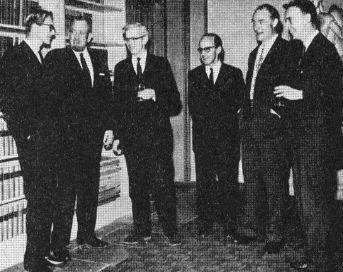
DNA Nobel Prize Celebration 1953
Rosalind Franklin (1920-1958), a graduate in physical chemistry, who helped found the science of high strength carbon fibres that found application as graphite rods in nuclear power plants, had been working since 1947 on x-ray crystallographic methods at the Central State Chemistry Labs. in Paris. As an expert on x-ray crystallography she was head hunted in 1951 by John Randall, of the Medical Research Council Biophysics Research Unit at King’s college, London, to work on an x-ray picture of DNA taken by a graduate student Raymond Gosling. The working relationship at King’s college did not get off to a good start because of a misunderstanding. Maurice Wilkins (1916- ), a physics graduate and member of the Medical Research Council Biophysics Research Unit who had been working on nucleic acids and x-ray diffraction of DNA, was away on holiday when Franklin arrived. Gosling, his second in command, stood in for him at the first office meeting and, since no work had been done on DNA for several months, it was all given to Franklin. Franklin was the kind of person that once they were given something to solve they kept it to themselves until it was complete. Not the way that Wilkins liked to run the lab at King’s college. When Wilkins returned he thought that Franklin was a high class technical assistant who was to supply the team with experimental data for it to analyse. This was not to be. Relationships were strained between Wilkins and Franklin, whereas Gosling got on with them both. Thus the seeds were sown for a rough ride for all concerned.
To solve the structure of DNA four ideas had to come together. The first was that the phosphate backbone was on the outside, bases on the inside. That the molecule was a double helix. That the strands were antiparallel. That it had a specific base pairing.
Franklin soon found out that by bundling super thin strands of DNA and zapping them with a super fine x-ray beam there were two forms of hydration — the A form (easy to photograph) that is dry and the B form (hard to photograph) that is wet. Her B form photographs showed a fuzzy cross which meant a helix. Since the water would be attracted to the phosphates in the backbone, and the DNA was easily hydrated and dehydrated, she guessed that the backbone was on the outside and the bases were on the inside. The first part of the problem was solved.
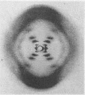
Photo 51 x-ray diffration of wet DNA
showing the B form double helix
taken by Rosalind Franklin
In the November of 1951 Franklin gave a departmental seminar to bring the unit up to date on what she had achieved so far. In it she presented the A and B form data. In the audience was an American James Watson (1928- ), a zoology graduate, who had become interested in molecular biology, with particular emphasis on DNA. He was on a National Foundation of Infantile Paralysis Fellowship at the University of Cambridge, England, and was working under Laurence Bragg and his x-ray team. After the seminar Watson caught the train back to Cambridge and based on what he had heard and seen he and Francis Crick (1916- ) built their first model of DNA. This was slightly unethical as Bragg’s Cambridge team had a gentleman’s agreement that they would work on x-ray crystallography of protein and Randall’s team in London would work on DNA. Watson had not taken notes, knew nothing about crystallography, and had only remembered bits of Franklin’s talk. So when Watson and Crick invited Franklin and Wilkins to view the model Franklin tore its construction to shreds. The model was a triple helix with bases on the outside. Egos wounded Bragg banned Crick and Watson from working on DNA.
In May, 1952 Franklin got the first good photograph of the B form of DNA. The photograph showed a double helix which was the second part of the problem that had to be proved. Franklin though was a perfectionist when it came to research and would not release any data until she had more information on the A form which was giving the most data.
Meanwhile in America Linus Pauling (1901-1994), a physical chemist with an interest in biological chemistry, who in 1950 constructed the first satisfactory model of a protein molecule and a twice winner of the Noble prize (1954 chemistry and 1962 Peace), was working on the structure of DNA and was very close to solving the puzzle. In May, 1952 he applied for a passport to visit England for a conference, but it was refused because of accusations of being a communist. Had he attended the conference he would almost certainly have met Franklin and had they collaborated with her data and his knowledge they would almost certainly have solved the structure of DNA.
Not releasing the information on the B form proved to be Franklin’s downfall for she got bogged down with calculations and obsessed in trying to determine whether the A form was helical. Gosling got so frustrated that on Friday the 18th of July, 1952 he announced on a blackboard the “Death of the A-Helix”. Franklin continued to waste most of the winter of 1952 with work on the A form. She did meet with Crick who tried to offer advice, but since his character was typically patronising she rejected an opportunity to collaborate. An opportunity missed for they would most certainly have solved the puzzle in months.
Meanwhile because of lack of communication at the King’s College lab Wilkins was trying to reproduce Franklin’s results, but he failed to get the same quality. Back in Cambridge Watson and Crick were working with a lot of volatile enthusiasm, but no data.
Early in 1953 Linus Pauling sent his son Peter, who was studying at Cambridge, a draft copy of his paper on DNA for comment. Watson knew Peter and somehow got a sight of the DNA paper. Watson took the paper to Franklin for comment, but she dismissed it as rubbish, being further annoyed as she had written to Pauling for information, but none had been sent. Upset at his reception Watson went to talk to Wilkins. Wilkins showed Watson one of Franklin’s B form photographs without her consent or knowledge. Then in February Max Perutz, head of the Medical Research Council Unit housed at the Cavendish Laboratory in Cambridge where Crick was a research student, received a government report that contained the data presented by Franklin at her departmental seminar. The report was not confidential, but it was private. Perutz passed the report on to Crick without asking Randall or Franklin’s permission.
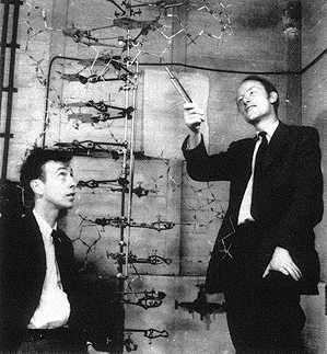
Francis Crick shows James Watson the model of DNA
in their room number 103 at the Cavendish Laboratories, Cambridge
Watson and Crick now had all Franklin’s data that showed that DNA was a multiple helix. Crick who had worked on proteins soon realised that Franklin’s data implied an antiparallel double helix. On the 10th of February Franklin began working on the B form again. According to Franklin’s note book she had had the antiparallel idea for the A form, but had not applied it to the B form, had she done so she would have solved part three of the puzzle earlier.
The last part of the puzzle focused on the base pairing and required a thorough knowledge of Chargaff’s rules. Watson and Crick had come to an elementary understanding of Chargaff’s rules the year before, with Crick even arranging a meeting with Chargaff where he had to admit that he did not even understand the basics let alone the rules. Franklin was conversant with Chargaff’s rules and so with all the facts at her fingertips and her superior knowledge of chemistry it was only a matter of time before she used logic to figure out the last part of the puzzle. In fact she had a draft paper dated the 17th of March, 1953 which outlined that the molecule was a double helix, had specific base pairing and the antiparallel A form which had not been applied to the B form. Franklin did not realise that Watson and Crick were racing to publish first, which they did on the 18th of March, 1953, so beating her because she had not published.
The Watson and Crick “Nature” paper cites no authorities or historical record. It contains no experimental proofs. It contains only hypotheses. No acknowledgement is made to Franklin and Wilkins at King’s college, London beyond the following statement. “We have also been stimulated by a knowledge of the general nature of the unpublished results and ideas of Dr. M.H.F. Wilkins, Dr. R.E. Franklin, and their co-workers at King’s College London.”.
In 1962 Watson, Crick and Wilkins received the Nobel Prize in Physiology or Medicine. They proposed that DNA molecule takes the shape of a double helix, an elegantly simple structure that resembles a gently twisted ladder. The rails of the ladder are made of alternating units of phosphate and the sugar deoxyribose; the rungs are each composed of a pair of nitrogen-containing nucleotides. In their Nobel lectures they cite 98 references, none are Franklin’s. Only Wilkins included her in his acknowledgements. Franklin died in 1958. The Noble Prize is only awarded to living persons.
The recipe for making DNA
Most people think that DNA is some kind of mysterious substance buried away in our cells never to be seen — but that is not the case. Here is a recipe for the extraction of DNA to make in your own kitchen — not unlike the type of experiment Rosalind Franklin would have performed in her lab at King’s college, London. She had in fact tried oranges which were not at all suited to this form of experiment and proved useless. Kiwi fruit is best suited to making DNA because of its structure.
You will need:
1. A ripe Kiwi fruit.
2. Table salt.
3. Non concentrated washing up liquid.
4. Knife and a chopping board.
5. Measuring scales.
6. A measuring jug.
7. A teaspoon (5 ml).
8. Small bowl.
9. A large sauce pan full of hot water (not boiling — about 60 degrees).
10. A large sauce pan full of ice.
11. Coffee filter and filter funnel.
12. A tall, thin plain glass.
13. Some thin wire (fuse wire).
What to do:
1. Put the bottle of methylated spirits in the ice.
2. Make up a solution of 3 grams of salt, 10 ml of washing up
liquid and 100ml of water. Measure 3 grams of salt .
You may find that your scales will weigh out a minimum of 25 grams. If this is
the case add 80 ml of washing up liquid (16 measuring spoons) and make
up to just less than one litre. Stir your mixture thoroughly (avoid froth) to
dissolve the salt.
3. Peel and chop one Kiwi fruit. Scoop this into the small bowl and add
100 ml of the salt — detergent mix.
4. Put the bowl containing the mixture into the saucepan of hot water and leave for 15 minutes.
5. After 15 minutes pour the mixture into the coffee filter, and catch the liquid
that filters through in the tall glass. You will need about one fifth of a glass.
6. Very carefully drizzle the ice cold meths down the inside of the
glass so that it forms a purple layer on top of the green layer.
When there is about a fifth of a glass of meths (i.e. two fifths including the
green liquid) put the glass on the table and watch what happens.
7. After a few moments you should see a white layer beginning to
form at the boundary between the green and the purple layers. If you have
done it correctly you will see that the layer is made up of a filament — DNA
from Kiwi fruit.
8. Try scooping the DNA out with a loop of thin wire — purple white goo - the
molecules of life.
What is happening:
DNA is found in every cell of every living thing, but it is difficult to get it out
and disentangle the DNA from the protein inside the cells.
The composition of Kiwi fruit allows the DNA to be extracted without a great
deal of effort, but not all things do. By chopping the Kiwi fruit and letting it
soak in the detergent and salt the first part of the problem is solved. The
detergent strips away the cell membranes that cover the inside of the cell
and lets the internal goo escape. The kiwi fruit needs to be chopped fine
enough to get the cells broken open, but this should not be overdone or the
DNA will be smashed.
The second part of the problem, getting rid of the protein that has stuck to the
DNA is all done by the Kiwi fruit. The Kiwi fruit already contains a lot of a
special enzyme called a proteinase. Enzymes are like small molecular
machines that do many things — the enzymes in washing powder
digest fat for example. The proteinase enzyme attacks the proteins clinging
to the DNA and breaks them up thereby releasing the DNA. Rosalind
Franklin tried oranges, but oranges do not contain enzymes and therefore
she failed to make DNA from them. She and Raymond Gosling finished up
having a food fight with the residue of their efforts.
The green layer that is produced in the glass is full of DNA as well as lots of
broken up proteins and other things. When the cold meths is poured onto the
green layer, the DNA dissolved in the water layer at the bottom of the glass is
turned into a solid as the DNA cannot remain in the solution. Little bubbles
may form between the two layers and drag the strands of DNA up into
the meths — the bubbles may be caused by the temperature difference
between the layers, making the air dissolved in the green layer come out of
the solution. © BA
The Original Report
We hope that this information has been useful to you.
To return to an index click its button below or the hat at the top of the page.